 Glaucoma is a leading cause of blindness in the United States. This eye disease, which is especially common after the age of 40, gradually degrades the optic nerve, eventually leading to partial or complete vision loss. The damage caused by glaucoma is not reversible, but with early treatment disease progression can be slowed so that individuals are able to preserve clear vision.
Glaucoma testing is the most essential tool in disease diagnosis and vision preservation. Individuals in the Harlingen, TX, area who have experienced symptoms of glaucoma can learn what to expect from a typical glaucoma exam with Dr. Raul Peña.
Comprehensive Eye Exam
A comprehensive eye exam is usually the first step in diagnosing glaucoma. Many people think a routine eye exam is all about testing the vision, but Dr. Peña looks at many other factors as well. Included in a comprehensive eye exam is a non-contact tonometry test. This exam, in which a light puff of air is directed at the eye, measures IOP, or intraocular pressure. Increased IOP is a potential sign of glaucoma. If our Harlingen patients have a higher IOP than expected, we will likely schedule a follow-up glaucoma exam, which will include multiple forms of testing.
Eye Pressure Check
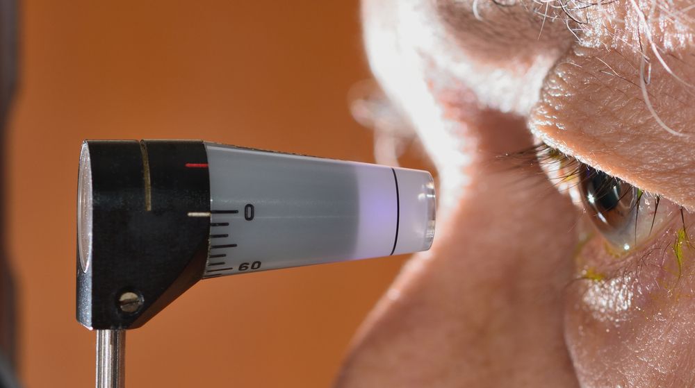
A glaucoma eye exam always includes an eye pressure check. The eye pressure exam performed for glaucoma diagnosis is more accurate than the non-contact tonometry test that is performed during a comprehensive eye exam. During this test, numbing eye drops are applied to desensitize the eye. A small instrument is then placed on the surface of the eye. The procedure is quick and painless, but it is important that patients remain relaxed, so that the inner eye pressure can be accurately measured.
Glaucoma Imaging Tests
Glaucoma imaging tests allow Dr. Peña to photograph the optic nerve and look for any signs of damage. This test requires the patient’s eyes to be dilated. Once the eyes are dilated the optic nerve is photographed using a digital camera or other optic nerve imaging techniques. The images are used to assess the shape, color, size, and depth of the vessels in the optic nerve. These images are essential in determining if glaucoma has caused any damage, as well as tracking any future damage the disease may cause.
While the eyes are dilated for glaucoma imaging testing, Dr. Peña is likely to examine the central and peripheral retina as well. This provides further information that can be useful in glaucoma diagnosis.
Visual Field Test
If Dr. Peña suspects that one of his Harlingen patients has glaucoma, a visual field test will tell him if the disease has caused any vision loss. Visual field tests are completely pain-free and only take a few minutes per eye. If a patient is diagnosed with glaucoma, visual field tests will be repeated periodically so that we can track the progression of the disease and alter the patient’s treatment plan, as necessary, to preserve their vision.
Corneal Thickness Test
During a glaucoma exam it is important to measure corneal thickness. An especially thin cornea may cause low eye pressure readings, while a thicker cornea could contribute to higher IOP readings. To measure corneal thickness the eye is numbed and a small probe is gently applied to the cornea.
Angle Test
The angle test is another exam that can be crucial in glaucoma diagnosis and treatment, because it lets Dr. Peña determine if the patient is suffering from open- or closed-angle glaucoma. Like many others, this test is performed while the eye is numb. During the test a gonioscopy lens (similar to a contact lens) is placed on the surface of the cornea. The gonioscopy lens makes the angle where the cornea meets the iris visible to Dr. Peña.
Learn More
Glaucoma testing is the only way to accurately diagnose glaucoma so that patients can be treated and the vision can be preserved. If you have more questions about what to expect during a glaucoma exam, Dr. Raul Peña can provide you with answers. To get in touch, send us a message online, or call our office at (956) 264-1200.

